Forschung
Clinical Neuroimaging
Clinical neuroimaging focuses on aspects of brain functioning that are relevant to the psychiatric clinic and may be important for future translation of this kind of knowledge into clinical practice.
Notwithstanding the enormous increase in knowledge brought about by the last 15-20 years of functional and structural neuroimaging studies, their translational impact on psychiatric practice has been modest. Translational neuroimaging may require rethinking the way we analyse data and develop specific statistical approaches (as opposed to applying techniques developed to localize functional activations). We focus on the identification of the neural substrates of individual differences that may be of relevance for mental health, as those emerging from genetic polymorphisms or individual differences in social cognition and decision making, and on innovative models of emotion regulation.
Key publications
- Messina, I., Grecucci, A., & Viviani, R. (2021) Neurobiological models of emotion regulation: A meta-analysis of neuroimaging studies of acceptance as an emotion regulation strategy. Cogn. Aff. Neurosci., doi: 10.1093/scan/nsab007.
- Sosic-Vasic, Z., Eberhard, J., Bosch, J.E., Dommes, L., Labek, K., Buchheim, A., & Viviani, R. (2019) Mirror neuron activations in encoding of psychic pain in broderline personality disorder. NeuroImage Clin., 22, 101737.
- Coch, C., Viviani, R., Breitfeld, J., Münzer, K., Dassler-Plencker, J., Holdenrieder, S., Coenen, M., Steffens, M., Müller, M., Hartmann, G., & Stingl, J. (2019) Interferon-beta-induced changes in neuroimaging phenotypes of appetitive motivation and reactivity to emotional salience. NeuroImage Clin., 24, 102020. (shared first authorship)
- Messina, I., Sambin, M., Beschoner, P., & Viviani, R. (2016) Changing views of emotion regulation and neurobiological models of the mechanism of action of psychotherapy. Aff. Behav. Neurosci., 16, 571-587.
- Messina, I., Sambin, M., Palmieri, A., & Viviani, R. (2013) Neural correlates of psychotherapy in anxiety and depression: A meta-analysis. PLoS One, doi: 10.1371/journal.pone.0074657.
- Viviani, R., Sim, E.J., Lo, H., Beschoner, P., Osterfeld, N., Maier, C., Seeringer, A., Godoy, A., Rosa, A., Comas, D., & Kirchheiner, J. (2010) Baseline brain perfusion and the serotonin transporter promoter polymorphism. Biol. Psychiatry, 67, 317-322.
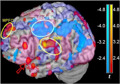
Neuroimaging of emotion and social cognition
We focus on spontaneous forms of emotion regulation and decision making in social cognition. The link between these two areas is the role of highly connected associating areas in determining recruitment of relevant schemas.
Key publications
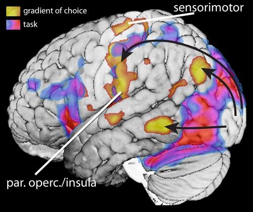
- Labek, K., Sittenberger, E., Kienhöfer, V., Rabl, L., Messina, L., Schurz, M., Stingl, J.C., & Viviani, R. (2021) The gradient model of brain organization in decisions involving 'empathy for pain'. bioRxiv, https://doi.org/10.1101/2021.11.28.470235
- Viviani, R., Dommes, L., Bosch, J.E., & Labek, K. (2020) Segregation, connectivity, and gradients of deactivation in neural correlates of evidence in social decision making. NeuroImage, 223, 117339.
- Viviani, R., Dommes, L., Bosch, J., Steffens, M., Paul, A., Schneider, K.L., Stingl, J.C., & Beschoner, P. (2020) Signals of anticipation of reward and of mean reward rates in the human brain. Reports, 10, 4287.
- Messina, I., Bianco, S., Sambin, M., & Viviani, R. (2015) Executive and semantic processes in reappraisal of negative stimuli: Insights from a meta-analysis of neuroimaging studies. Frontiers Psychol., doi: 10.3389/fpsyg.2015.00956.
- Viviani, R. (2014) Neural correlates of emotion regulation in the ventral prefrontal cortex and the encoding of subjective value and economic utility. Frontiers Psychiatry, doi: 10.3389/fpsyt.2014.00123.
- Viviani, R., Lo, H., Sim, E.J., Beschoner, P., Stingl, J.C., & Horn, A.B. (2010) The neural substrate of positive bias in spontaneous emotional processing. PLoS One, 5, e15454.
Genetic and pharmacogenetic imaging
Some drug-metabolizing enzymes, such as CYP2D6, are also expressed in the brain. Our studies investigate neural correlates of genetic polymorphisms of these enzymes
Key publications
- Stingl, J.C., Scholl, C., Bosch, J.E., & Viviani, R. (2021) Genetic polymorphism of CYP2C19 and subcortical variability in the human adult brain. Psychiatry, 11, 467.
- Viviani, R., Messina, I., Bosch, J.E., Dommes, L., Paul, A., Schneider, K.L., Scholl, C., & Stingl, J.C. (2020) Effects of genetic variability of CYP2D6 on neural substrates of sustained attention during on-task activity. Psychiatry, 10, 338.
- Viviani, R., Abler, B., Seeringer, A., & Stingl, J.C. (2012) Effect of paroxetine and bupropion on human resting brain perfusion: An arterial spin labelling study. NeuroImage, 61, 773-779.
- Stingl, J.C., Brockmöller, J., & Viviani, R. (2013) Genetic variability of drug-metabolizing enzymes: The dual impact on psychiatric therapy and regulation of brain function. Psychiatry, 18, 273-287.
- Kirchheiner, J., Seeringer, A., Godoy, A.L., Maier, C., Beschoner, P., Sim, E.J., & Viviani, R. (2011) CYP2D6 in the brain: Genotype effects on resting brain perfusion. Psychiatry, 16, 333-341.
- Viviani, R., Sim, E.J., Lo, H., Beschoner, P., Osterfeld, N., Maier, C., Seeringer, A., Godoy, A., Rosa, A., Comas, D., & Kirchheiner, J. (2010) Baseline brain perfusion and the serotonin transporter promoter polymorphism. Psychiatry, 67, 317-322.
Statistical analysis of imaging data
The statistical analysis of neuroimaging data is a challenging task. Typical imaging data cover the brain using over 200.000 voxels, whose activity is reciprocally related in complex ways. Dimensionality reduction and sophisticated forms of eigendecomposition of multimodal data may play an increasing important role in the future.
Key publications
- Viviani, R. (2021) Overcoming bias in representational similarity analysis. arXiv (preprint), 2102.08931.
- Viviani, R., Stöcker, T., & Stingl, J.C. (2017) Multimodal FLAIR/MPRAGE segmentation of cerebral cortex and cortical myelin. NeuroImage, 152, 130-141.
- Bachmann, S., Haffer, S., Beschoner, P., & Viviani, R. (2011) Imputation techniques for the detection of microstructural changes in schizophrenia, with an application to magnetization transfer imaging. Res., 132, 92-96.. (IF: 4.374)
- Viviani, R. (2010) Unbiased ROI selection in neuroimaging studies of individual differences. NeuroImage, 50, 184-189.
- Viviani, R., Beschoner, P., Ehrhard, K., Schmitz, B., & Thöne, J. (2007) Non-normality and transformations of random fields, with an application to voxel-based morphometry. NeuroImage, 35, 121-130.
- Viviani, R., Grön, G., & Spitzer, M. (2005) Functional principal component analysis of fMRI data. Br. Mapping, 24, 109-129.
Digital atlas vessels
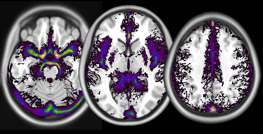
In the figure: a digital atlas of the frequency of occurrence of large vessels (arterial as well as venous), computed from a database of time-of-flight images, overlaid on a template structural image from the MNI. The digital atlas of vessels is freely available and can be found here:
https://identifiers.org/neurovault.collection:1061
This is a freely available digital atlas of frequency of vascularisation, obtained from N = 38 healthy participants scanned with the time-of-flight magnetic resonance technique. Images are available in NIfTI format in different voxel sizes:
Viviani, R. (2016) A digital atlas of middle to large brain vessels and their relation to cortical and subcortical structures. Frontiers Neuroanat., 10, 12.
The main source of information is the atlas in the original high resolution. The volume contains the frequency of detection of vessels in our dataset, in percent, and may be used to visualize their course by overlaying it together with activation maps. Volumes are also provided after resampling at the isotropic resolutions of 1 and 2mm, which are commonly used in structural or functional studies. While the incidence of vessels in the low-resolution atlas is not quantitatively identical due to the increased influence of partial volume effects, the location of vascular tissue corresponds closely to that displayed in the high-resolution atlas. These volumes may be used to create masks staking out regions where the vascular density is high.
When using these data to interpret findings of other studies, it should be remembered that the quality of spatial correspondence is limited by possible difficulties in redressing differences in brain shape induced by the geometry of the magnetic field of the original volumes, by the signal loss due to different susceptibility artefacts, and the possible use of customized normalization templates across specific studies and human populations. To enable users to assess and potentially address these discrepancies, the mean TOF normalized volume from all subjects in the study is provided. This volume contains structural information that may be used to compute the cross-normalization to another template.