Electron Microscopy
Contact: elmi-zoology@uibk.ac.at
History
Electron microscopy has a very long tradition at the Zoological Institute at the University of Innsbruck. Already in 1959 a so called Siemens Elmiskop 1 was installed and it was operative until 1986. In 1987 a Zeiss TEM 902 equipped with a prism-mirror-prism spectrometer was purchased. This allowed element analysis with EELS (Electron Energy Loss Spectroscopy) and it produced images with high contrast using a slit aperture to eliminate inelastic scattered electrons. In 2005 the Zeiss 902 was replaced with a fully digital Zeiss Libra 120 Energy Filter Transmission Microscope (EFTEM).
At present the Institute of Zoology provides two electron microscopes: A Zeiss Libra 120 Energy Filter Transmission Electron Microscope and a Zeiss DSM 950 Scanning Electron Microscope.
EFTEM Zeiss Libra 120
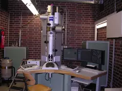
The in-column omega filter allows the elimination of inelastically scattered electrons. In combination with a slit aperture it produces routinely brilliant images with high contrast. The imaging of inelastically scattered electrons makes element analysis with EELS possible and shows the element distribution by using ESI (Electron Spectroscopic Imaging). Even HCI (High Contrast Imaging) of unstained specimens or the imaging of thick sections is now possible in routine work. This microscope is fitted also with a 5-axes goniometer with a 70° tilt angle.
Electron Energy Loss Spectroscopy (EELS)
The event of energy loss is produced by an electron coming from the electron beam which collides with an orbital electron of an atom of the specimen. The collision scatters this electron inelastically and the energy loss of the electron is specific for the element. A spectrometer can measure the element specific energy loss in a range from 0 to 2500 eV with a resolution of 1.5 eV.
Energy Spectroscopic Imaging (ESI)
For imaging of elements in a sample one image is produced at the edge of the energy loss of the particular element. In addition two images are produced below the edge of the element to reduce background and an HCI image completes the series. Subtraction of the three images shows the net distribution of the element. The element distribution in false colours is superimposed over the High Contrast Image which provides information on the ultrastructure of the tissue.
High Contrast Imaging (HCI)
To obtain high contrast images of unstained specimens an electron loss of 250 eV near the carbon edge is used. The result is a negative image with high contrast which then is inverted by the imaging software.
Members of this workgroup are:
- Anna Seybold